Skip to Content

INTRAVASCULAR IMAGING

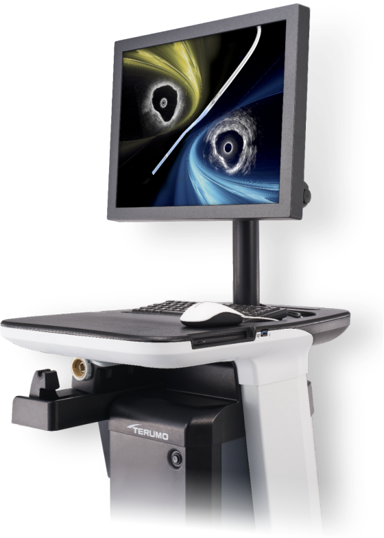
OPUSWAVE™ Imaging System

Simultaneous IVUS and OFDI System

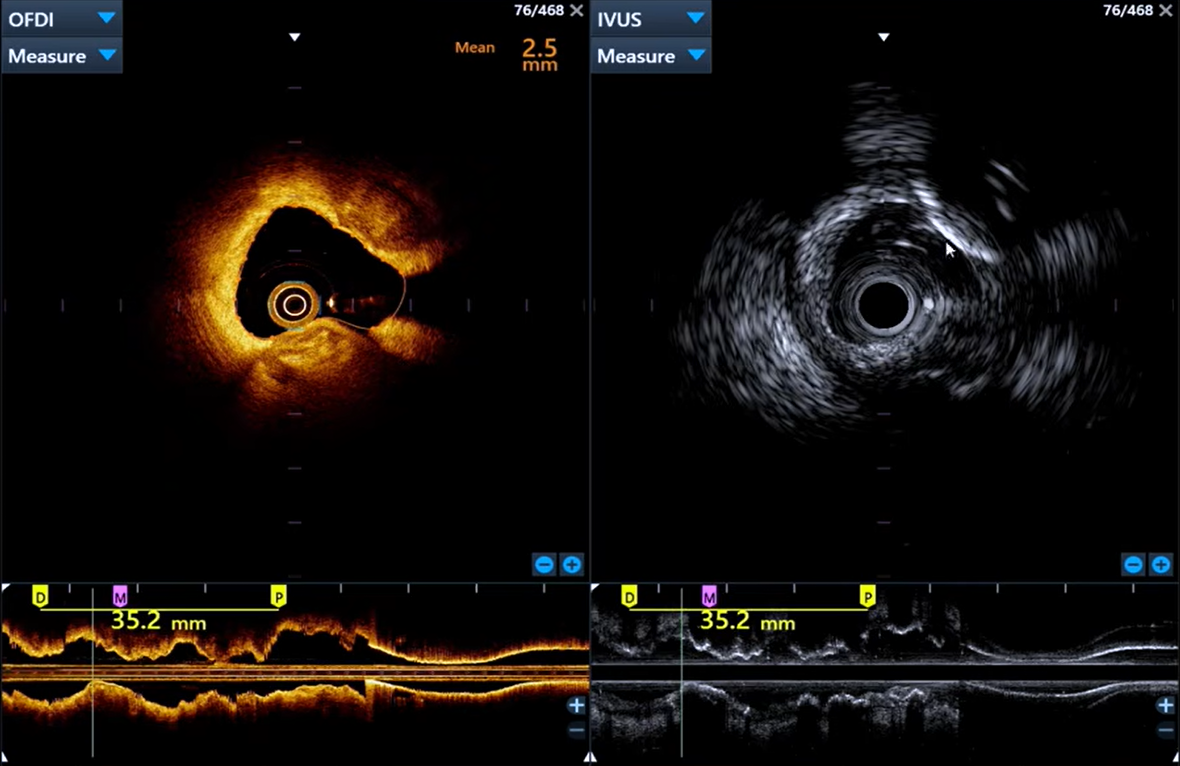
The Dual Sensor System Imaging Platform sets a new standard in intravascular imaging, delivering simultaneous IVUS and OFDI (next-generation OCT) modalities on a single screen. Capture both deep vessel structure and high-resolution surface detail in one pullback,providing a comprehensive view of the vessel.1

Product photo: OPUSWAVE Imaging System

Dual View. Single Clarity.

  • The OPUSWAVE Dual Sensor System delivers simultaneous IVUS and OFDI imaging to reveal high resolution surface detail and deep tissue penetration in a single pullback, enabling comprehensive lesion assessment.1

Flexibility at Your Fingertips.

  • Flexible display options allow clinicians to selectively choose IVUS or OFDI when every decision matters.

Dual Sensor, 2.6Fr Catheter.

  • Featuring advanced hydrophilic coating and 150mm pullback.

DUALVIEW™ Imaging Catheter

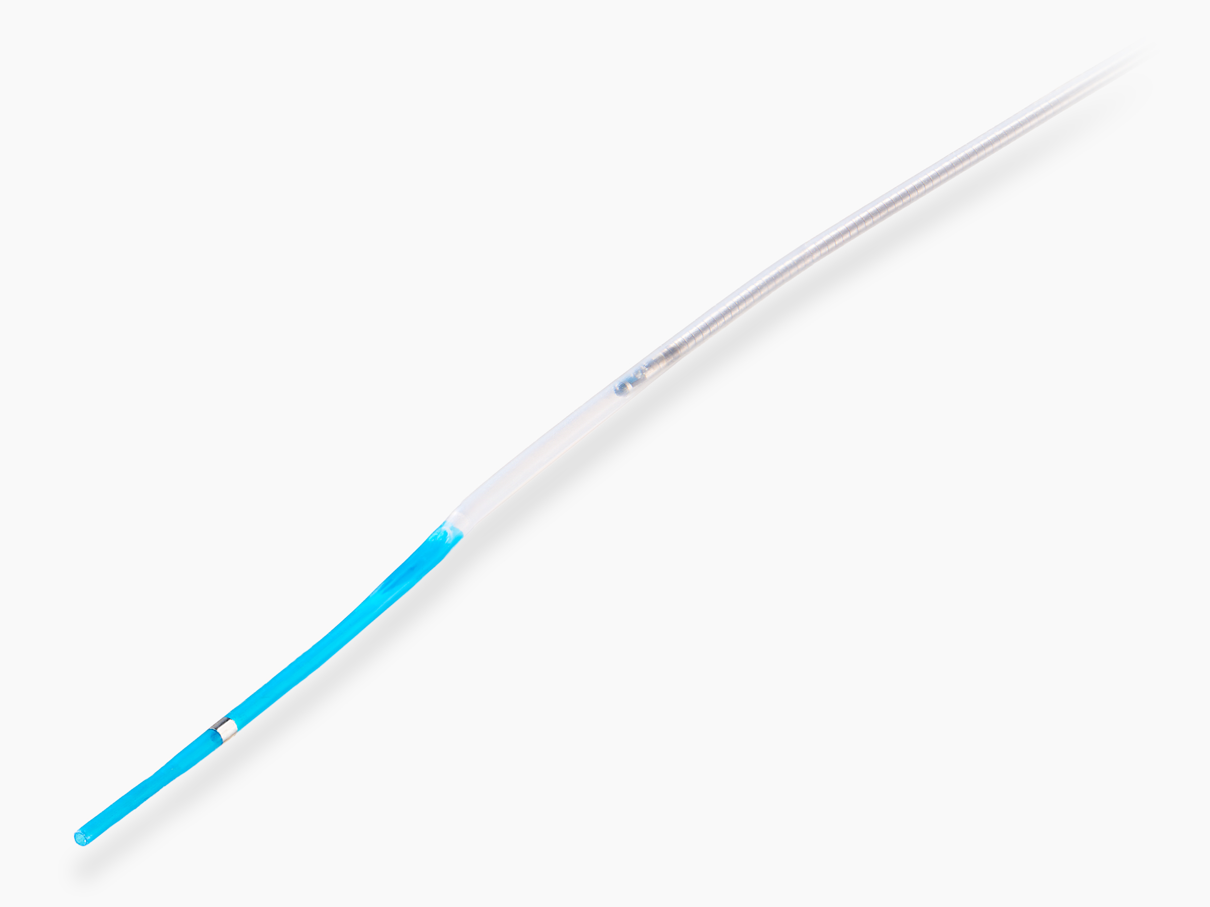
Terumo’s dual-sensor 2.6Fr catheter combines deliverability and a 150mm controlled pullback to keep you in position while you choose IVUS, OFDI, or Dual View. The DUALVIEW catheter features an advanced hydrophilic coating that becomes lubricious on contact with saline or blood, helping reduce vessel friction for enhanced trackability and deliverability.

Product photo: DUALVIEW™ Imaging Catheter

Additional Resources

The Terumo Dual Sensor System (DSS), with the OPUSWAVE Imaging Console and DUALVIEW catheter, was developed to provide both IVUS and OFDI images in a single pullback.  With their inherent strengths and limitations, the combined IVUS and OFDI probes are complementary and work synergistically to enable a comprehensive depiction of coronary arteries.1 

Intravascular Ultrasound and Optical Coherent Tomography Combined Catheter (2023)2

As IVUS and OCT have evolved in parallel over the years, hybrid IVUS-OCT systems have also been developed to merge the advantages of both modalities into a single catheter.  This review aims to summarize the differences and complementary aspects of IVUS and OCT and describe the novel hybrid IVUS–OCT catheter systems.

Advances in IVUS/OCT and Future Clinical Perspective of Novel Hybrid Catheter System in Coronary Imaging (2020)3

This review highlights how the complementary strengths of IVUS and OCT can provide a more complete understanding of coronary anatomy, plaque morphology, and procedural results. The authors suggest that hybrid imaging systems represent the next evolution in coronary imaging, offering comprehensive insights into vessel morphology.

2025 ACC/AHA/ACEP/NAEMSP/SCAI Guideline for the Management of Patients With Acute Coronary Syndromes: A Report of the American College of Cardiology/American Heart Association Joint Committee on Clinical Practice Guidelines4

Use of Intracoronary Imaging – Updated Class of Recommendation and Level of Evidence to 1A in patients with ACS undergoing coronary stent implantation in the left main artery or in complex lesions, intracoronary imaging with intravascular ultrasound (IVUS) or optical coherence tomography (OCT) is recommended for procedural guidance to reduce ischemic events. 

Expert Consensus Statement for Quantitative Measurement and Morphologic Assessment of Optical Coherence Tomography: Update 20255

In this updated expert consensus document, the methods for the quantitative measurement and morphologic assessment of optical coherence tomography (OCT) / optical frequency domain imaging images (OFDI) are briefly summarized. The focus is on the clinical application and the clinical evidence of OCT / OFDI to guide percutaneous coronary interventions.

Intravascular Imaging vs Angiography Guidance for PCI of Severely Calcified Lesions: The ECLIPSE Trial (2025)6

The aim of this study was to determine, from a large-scale randomized trial of PCI in severely calcified lesions, whether IVI guidance improves event-free survival. The primary clinical outcome was the 1-year rate of target vessel failure (TVF).

Intravascular Imaging-Guided Coronary Drug-Eluting Stent Implantation: An Updated Network Meta-Analysis (2024)7

Compared with angiography guidance, intravascular imaging guidance for coronary stent implantation with OCT or intravascular ultrasound enhances both the safety and effectiveness of PCI, reducing the risks of death, myocardial infarction, repeat revascularization, and stent thrombosis.

IVUS-Guided Versus OCT-Guided Coronary Stent Implantation: A Critical Appraisal (2018)8

 The purpose of this review is to describe the similarities and differences of IVUS and OCT, and to highlight the evidence supporting the utility of each in patients undergoing PCI with stent implantation. Optical coherence tomography (OCT) provides similar information to IVUS (with some important differences) and may also improve acute and long-term patient outcomes compared to angiography-guided PCI.

Preparing the OPUSWAVE MDU

Preparing the DUALVIEW Imaging Catheter

Connecting the DUALVIEW Catheter to the OPUSWAVE MDU

DUALVIEW Catheter Calibration

Performing a Pullback With the OPUSWAVE MDU

Reviewing OPUSWAVE Captured Images

Conducting Image Measurements With OPUSWAVE

OPUSWAVE Procedure Conclusion and File Management


OPUSWAVE Imaging System

PRODUCT CODE  IMAGING MODES  CO-REG.
01000-00-US Frame Rate: Dual Mode 100-160 fps
IVUS Only 30-100 fps
x
Pullback Speed: Dual Mode - 10, 20, 30, 40 mm/sec
IVUS Only - 0.5, 1, 2, 3, 10
x

DUALVIEW Imaging Catheter

PRODUCT CODE     
TU-DSS01C Imaging Window Profile  2.6F
Guidewire Rapid Exchange Rail Length  20mm
Transducer Frequency: IVUS mode  40 MHz
Guide Catheter Compatibility  6F
Guidewire Maximum OD  0.014”
Max. Pullback Length  150mm
Distal Radiopaque Marker to Tip  7mm
Distal Tip to Sensor  25mm
Distal Depth Markers 90cm, 100cm
Effective Length 137cm
Distal Hydrophilic Coating Length 100cm
Infrared Light for OCT Imaging  1300nm
INJECTOR SETTINGS
Flush Rate 4 mL/s
Total Volume 16mL
Pressure Limit 300 psi
(2068 kPa)

How do IVUS and OFDI dual-sensor catheters work together?

IVUS and OFDI dual-sensor catheters combine two complementary imaging technologies into a single platform. IVUS provides deep vessel penetration, enabling assessment of vessel size, plaque burden, and overall anatomy, while OFDI delivers near-microscopic resolution for detailed evaluation of plaque characteristics, calcium morphology, stent apposition, and procedural outcomes. By acquiring co-registered images simultaneously, the dual-sensor system allows physicians to visualize both the vessel's full structure and its fine luminal details in a single pullback.

What is the potential clinical advantage of using combined IVUS and OFDI during a PCI?

Combined IVUS and OFDI have the potential to provide both comprehensive vessel assessment and high-resolution lesion visualization from a single imaging platform. IVUS can provide information regarding vessel size, plaque burden, and lesion length, while OFDI can offer detailed visualization of features such as calcium morphology, plaque microstructure, stent expansion, malapposition, and edge dissections. By providing complementary imaging information, the combined use of IVUS and OFDI may support lesion assessment, procedural planning, device sizing, and post-stent evaluation. This integrated approach has the potential to provide clinicians with a more comprehensive view of coronary vessel and plaque morphology during PCI.

What are the pullback speeds and lengths of a DUALVIEW imaging catheter?

The DUALVIEW imaging catheter supports a maximum pullback length of 150 mm and variable pullback speeds of up to 40 mm/second in dual-imaging mode. This enables rapid acquisition of co-registered IVUS and OFDI images across long coronary segments, providing comprehensive vessel assessment and detailed lesion characterization during a single pullback.

Explain the technology behind Terumo's OPUSWAVE system.

OPUSWAVE is a dual-sensor intravascular imaging system that integrates Optical Frequency Domain Imaging (OFDI) and Intravascular Ultrasound (IVUS) within a single catheter. The DUALVIEW catheter houses both optical and ultrasound imaging elements, enabling simultaneous acquisition of co-registered OFDI and IVUS datasets during a single pullback. OFDI generates high-resolution cross-sectional images using near-infrared light and interferometric signal processing, while IVUS uses reflected ultrasound waves to image deeper vessel structures. The system synchronizes and displays both image streams in real time, allowing visualization of corresponding vessel segments from the two modalities at the same longitudinal location.

What are the latest innovations in coronary artery disease mapping?

Recent innovations in coronary artery disease mapping are centered on multimodality imaging, physiologic assessment, and advanced image integration. The most notable development is the emergence of dual-sensor intravascular imaging platforms that combine IVUS and OFDI within a single catheter, enabling simultaneous visualization of vessel architecture and plaque microstructure. Additional advances include AI-assisted plaque characterization, co-registration of physiologic measurements such as FFR and iFR with intravascular imaging, and three-dimensional vessel reconstruction. Together, these technologies provide increasingly detailed spatial characterization of coronary anatomy, plaque morphology, lesion severity, and stent geometry during PCI.

References

  1. Data on file
  2. Shigetaka Kageyama, Nozomi Kotoku, Kai Ninomiya, Shinichiro Masuda, Jiayue Huang, Takayuki Okamura, Scot Garg, Isao Mori, Brian Courtney, Faisal Sharif, Christos V. Bourantas, Patrick W. Serruys, Yoshinobu Onuma, Intravascular Ultrasound and Optical Coherent Tomography Combined Catheter, Interventional Cardiology Clinics, Volume 12, Issue 2, 2023, Pages 187-201, ISSN 2211-7458, ISBN 9780323939973, https://doi.org/10.1016/j.iccl.2022.12.002
  3. Ono M, Kawashima H, Hara H, Gao C, Wang R, Kogame N, Takahashi K, Chichareon P, Modolo R, Tomaniak M, Wykrzykowska JJ, Piek JJ, Mori I, Courtney BK, Wijns W, Sharif F, Bourantas C, Onuma Y and Serruys PW (2020) Advances in IVUS/OCT and Future Clinical Perspective of Novel Hybrid Catheter System in Coronary Imaging. Front. Cardiovasc. Med. 7:119. Doi: 10.3389/fcvm.2020.00119
  4. Rao, S, O’Donoghue, M, Ruel, M. et al. 2025 ACC/AHA/ACEP/NAEMSP/SCAI Guideline for the Management of Patients With Acute Coronary Syndromes: A Report of the American College of Cardiology/American Heart Association Joint Committee on Clinical Practice Guidelines. JACC. 2025 Jun, 85 (22) 2135–2237. https://doi.org/10.1016/j.jacc.2024.11.009
  5. Fujii, K., Kubo, T., Otake, H. et al. Expert consensus statement for quantitative measurement and morphologic assessment of optical coherence tomography: update 2025. Cardiovasc Interv and Ther 40, 226–233 (2025). https://doi.org/10.1007/s12928-024-01080-8
  6. Stone GW, Genereux P, Maehara A, Lewis BE, Shlofmitz RA, Dohad S, Choudary J, Dahle T, Pineda AM, Shunk K, Mahmud E, Weisz G, Collins MB, Alaswad K, Kumar G, Solankhi N, Dulas D, Altman JD, Kraemer C, Stiefel KM, Jones DE, Buccola JR, Kirtane AJ. Intravascular Imaging vs Angiography Guidance for PCI of Severely Calcified Lesions: The ECLIPSE Trial. JACC Cardiovasc Interv. 2025 Oct 13;18(19):2338-2351. Doi: 10.1016/j.jcin.2025.08.024. PMID: 41093451.
  7. Gregg W Stone, Evald H Christiansen, Ziad A Ali, Lene N Andreasen, Akiko Maehara, Yousif Ahmad, Ulf Landmesser, Niels R Holm, Intravascular imaging-guided coronary drug-eluting stent implantation: an updated network meta-analysis, The Lancet, Volume 403, Issue 10429, 2024, Pages 824-837, ISSN 0140-6736, https://doi.org/10.1016/S0140-6736(23)02454-6
  8. Maehara A, Matsumura M, Ali ZA, Mintz GS, Stone GW. IVUS-Guided Versus OCT-Guided Coronary Stent Implantation: A Critical Appraisal. JACC Cardiovasc Imaging. 2017 Dec;10(12):1487-1503. Doi: 10.1016/j.jcmg.2017.09.008. PMID: 29216976.

Image Detail: